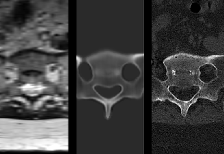
Spine surgery pre-operative planning has long suffered from a costly diagnostic fragmentation: magnetic resonance imaging (MRI) provides excellent visualization of soft tissues, spinal cord compression, and nerve root impingement, but lacks the radio-density characteristics required to visualize cortical bone morphology.
For some spine procedures, surgeons request CT as well as MRI to assess bone and plan instrumentation. An additional scan can add scheduling, cost and radiation exposure, although the need and delay vary by patient and institution. The FDA 510(k) clearance of MRI2CT's NextMR Spine platform fundamentally disrupts this paradigm by synthesizing high-precision 3D bone models directly from standard soft-tissue MRIs.
In This Deep Dive:
- Why this matters now: The clinical and economic friction of dual-modality spine imaging.
- What actually happened: NextMR Spine FDA 510(k) Class II clearance, 60-second processing, and submillimeter accuracy across 164 patients.
- The obvious read versus the deeper signal: Standalone SaMD viewer versus single-modality robotic navigation infrastructure.
- Competitive taxonomy & clinical maturity: Orthopedic synthetic imaging and 3D reconstruction platforms.
- The Evidence Ladder: Deep learning voxel conversion to multi-center surgical validation.
- The HealthTech Investor's Signal: Episode-of-care cost compression, ASC scheduling throughput, and MedTech OEM licensing.
- Counter-thesis: Complex anatomy, acquisition variability and implementation economics.
- Forward intelligence: 4 observable test indicators for the upcoming 12 to 24 months.
- The bottom line for orthopedic surgeons, hospital radiology chairs, and digital health venture investors.
Why this matters now
Lumbar fusion is a resource-intensive inpatient and ambulatory procedure. Precise pre-operative anatomical visualization is critical for selecting pedicle screw trajectories, planning interbody cage dimensions, and registering patient anatomy during robotic and navigation-assisted procedures.
Because MRI signals reflect proton density and tissue relaxation times rather than X-ray attenuation, cortical bone appears largely as a dark signal void on standard MRI sequences. Surgeons could not reliably distinguish fine bony landmarks, osteophytes, or facet joint arthrosis without ordering a high-resolution CT scan. By applying advanced deep generative neural networks to standard 1.5T and 3T MRI sequences, NextMR Spine generates synthetic CT (sCT) volumetric DICOM datasets that provide bone visualization; attenuation equivalence and geometric performance must be evaluated for the intended use, potentially reducing the need for a secondary CT in suitable cases, subject to clinician review and the cleared intended use.
What actually happened
On September 4, 2026, the FDA granted 510(k) clearance (K260525) to MRI2CT Inc. for NextMR Spine, a cloud-native radiological software application that automates the 3D reconstruction of bone morphology of the lumbar spine and sacrum from routine MRIs. The zero-footprint web application ingests standard clinical MRI protocols without requiring proprietary pulse sequences or specialized scanner hardware.
The FDA summary reports retrospective performance validation using 164 subjects. This supports the cleared imaging function but should not be described as a prospective surgical-outcomes trial. The algorithm automatically produces multiplanar reformats, 3D volumetric renders, and standard DICOM output files in under 60 seconds, supporting image export into compatible workflows; specific navigation integration requires its own validation.
The obvious read versus the deeper signal
The surface-level interpretation is that MRI2CT has created a convenient AI visualization tool for radiology viewing suites. The deeper structural signal is the establishment of single-modality orthopedic workflows that compress episode-of-care economics and could support future surgical planning integration.
In value-based orthopedic care and ambulatory surgery centers (ASCs), diagnostic delay directly impairs surgical conversion rates. Eliminating the pre-operative CT removes scheduling bottlenecks, reduces payer authorization hurdles, and avoids the radiation from a CT that is genuinely no longer required; dose varies with protocol. More strategically, synthetic CT serves as the foundational data layer for spine robotics OEMs (such as Medtronic Mazor, Globus Medical ExcelsiusGPS) to offer streamlined, MRI-based pre-planning, where appropriate; this does not establish clearance for autonomous robotic navigation or guarantee radiation-free surgery.