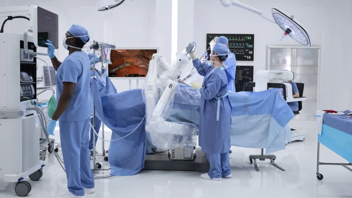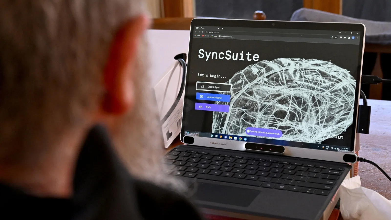
Inside Operating Room 4, a surgical team is preparing to resect a deep-seated glioma located near the motor strip. The patient is secured in a rigid skull clamp. The navigation system displays a three-dimensional model of the patient's brain, reconstructed from high-resolution magnetic resonance imaging acquired the previous evening. The system uses infrared cameras to track the surgeon's pointer relative to the patient's head, showing the tip of the instrument on the screen with sub-millimeter theoretical accuracy.
However, the moment the surgeon incises the dural membrane, the physical coordinate system changes. Cerebrospinal fluid drains from the ventricles, the tumor is partially debulked, and gravity pulls the hemisphere downward. The brain tissue visibly moves. This physical displacement, known as brain shift, can range from two to over ten millimeters during a standard craniotomy. When the target tissue is adjacent to critical pathways governing speech or motor function, a tracking error of even two millimeters can lead to permanent neurological deficits.
This mismatch represents the primary limitation of modern stereotactic navigation. While the tracking hardware is highly precise, the underlying map is static. The future of artificial intelligence in neurosurgery lies not in automating the surgeon's hands, but in transforming this static map into a dynamic, self-updating coordinate system that adapts to the moving anatomy of the brain. This approach, known as dynamic navigation, relies on constant sensor updates and predictive algorithms to keep the surgical map accurate throughout the procedure.
The Physics of Dynamic Navigation
To solve the challenge of anatomical movement, developers are focusing on algorithms capable of updating surgical plans in real time. Currently, correcting for tissue displacement requires pausing the operation to acquire intraoperative ultrasound scans or moving the patient into a specialized, high-cost intraoperative magnetic resonance imaging suite. Both methods introduce clinical delays and physical disruption to the surgical workflow.
The emerging computational alternative is deformable registration, which refers to the mathematical alignment of historical, high-resolution pre-operative scans with real-time, lower-resolution intraoperative data. This process relies on deep learning models trained on biomechanical simulations of brain tissue behavior. By analyzing how different regions of the brain deform under specific surgical positions, skull opening sizes, and gravity vectors, these networks can predict tissue movement.
When the surgeon acquires a rapid, localized intraoperative ultrasound, the artificial intelligence model does not simply display the raw, noisy ultrasound image. Instead, it uses the ultrasound data as anchor points to warp and correct the pre-operative magnetic resonance images. This provides the surgeon with an updated, high-fidelity map of the deep tracts and tumor boundaries without requiring an intraoperative magnetic resonance scan. For developers, the commercial opportunity lies in building software packages that integrate directly with existing optical tracking hardware, upgrading legacy navigation systems with real-time update capabilities.
Semantic Segmentation at the Microscope Eyepiece
The primary interface for a neurosurgeon during a microdissection is the surgical microscope. While heads-up displays have begun projecting simple navigation graphics into the eyepiece, these overlays are often distracting because they are not context-aware. They show static targets rather than understanding what the surgeon is actively looking at.
Computer vision models trained on thousands of hours of microsurgical video are changing this interface. By performing real-time semantic segmentation, these models can identify and color-code distinct anatomical structures directly within the optical path. The system can instantly distinguish between normal brain tissue, edema, active tumor margin, and critical vascular structures.
This capability is particularly valuable when paired with optical fluorescence. Surgeons often use specialized dyes like five-aminolevulinic acid, which causes high-grade glioma cells to glow red under blue light. However, the boundary between faint fluorescence and normal tissue is highly subjective and easily obscured by bleeding. Computer vision algorithms can analyze the pixel-level color variations under blue light far more precisely than the human eye, outlining the exact boundary of the tumor margin in real time. This precise margin identification helps maximize the extent of resection while sparing healthy tissue, which remains the single most important prognostic factor in neuro-oncology.
Preservation of the White Matter Architecture
Removing a tumor is only half the battle; the surgeon must also preserve the neural pathways that connect different regions of the brain. These pathways, or white matter tracts, are visualized using a technique called diffusion tensor imaging, which maps the direction of water diffusion along myelinated axons to reconstruct the brain's structural cabling.
Historically, tractography has been highly operator-dependent. A technician must manually select seed regions to generate the tract reconstructions, leading to high variability between institutions and even individual cases. Furthermore, current systems struggle to accurately map tracts that are compressed or distorted by a large tumor, often failing to show pathways that are physically present but functionally compromised.
Machine learning models trained on large datasets of brain connectivity are standardizing this process. These networks can automatically segment and reconstruct major tracts, such as the corticospinal tract and the arcuate fasciculus, with minimal manual input. More importantly, they can perform predictive reconstruction, estimating where a displaced tract should lie even when the local imaging signal is severely degraded by edema or tumor infiltration. By integrating these reconstructed tracts into the dynamic navigation system, the surgical planning software can warn the operator when an instrument is approaching a critical pathway, providing a digital guardrail during deep tissue dissection.
Closed-Loop Robotics and Smart Instruments
Beyond visualization, the integration of artificial intelligence with robotic hardware represents the next physical step in neurosurgery. While autonomous robotic surgery remains far in the future, passive and semi-active robotic assistance is already entering clinical use.
In these systems, the robot does not perform the cut. Instead, it holds the instrument guide with absolute stability, aligning itself automatically based on the surgical plan. For procedures like deep brain stimulation lead placement or stereotactic biopsies, where a needle must be guided to a precise target deep within the brain, robotic arms eliminate human tremor and mechanical drift.
The next phase of this technology involves smart instruments equipped with sensor arrays that feed data back to machine learning algorithms. For example, smart micro-forceps can measure mechanical resistance and electrical impedance at the tip of the tool. Since different tissue types, such as healthy white matter, scarred tissue, and tumor, exhibit distinct physical properties, the algorithm can analyze this feedback in real time. If the instrument detects that the surgeon is applying pressure to a structure with the physical signature of a major blood vessel, the system can increase haptic resistance in the robotic controller, physically preventing the surgeon from advancing further. This closed-loop feedback turns passive tools into active partners in the operating room.
Navigating the Regulatory and Data Bottlenecks
For healthtech operators and founders, the primary challenges to deploying these advanced tools are clinical validation and data access. Training robust computer vision and tissue deformation models requires vast amounts of high-quality, annotated intraoperative data. However, surgical videos and intraoperative imaging datasets are highly siloed within academic medical centers, and clinical annotation is time-consuming for practicing neurosurgeons.
Furthermore, the regulatory pathway for adaptive software is complex. The Food and Drug Administration has established frameworks for locked algorithms, but models that update their parameters or recommendations in real time present novel validation challenges. Startups must prove that their real-time registration algorithms do not introduce errors or artifacts that could mislead a surgeon.
To succeed, companies must design clinical studies that measure clear, objective endpoints, such as reduction in operative time, increased extent of tumor resection, or decreased rate of post-operative neurological deficits. Technology that merely adds complexity to the operating room without demonstrating measurable clinical or financial benefit will struggle to find adoption in an increasingly value-oriented healthcare system.
Key Signals
Real-time deformable registration software is transitioning from academic research to commercial integration, allowing existing optical hardware to correct for tissue movement without requiring costly intraoperative imaging.
Computer vision models integrated into the surgical microscope will soon provide active boundary delineation during tumor resections, shifting the interface from passive visual overlays to context-aware clinical assistance.
The successful scaling of AI in the neurosurgical suite depends on creating standardized clinical datasets that allow algorithms to be trained across diverse patient demographics and surgical approaches.