At 2:00 AM in a busy neuro-intensive care unit, a patient recovering from a ruptured aneurysm exhibits a subtle decline in neurological response. The standard clinical pathway requires immediate imaging to rule out vasospasm or hydrocephalus. In most hospitals, this means organizing an intra-hospital transport to the radiology department, a process requiring an ICU nurse, a respiratory therapist, and a transport technician to manage the patient's ventilator, intravenous infusions, and arterial lines. The physical movement of such critically ill patients carries a documented adverse event rate of up to fifteen percent, ranging from accidental extubation to transient hypotension.
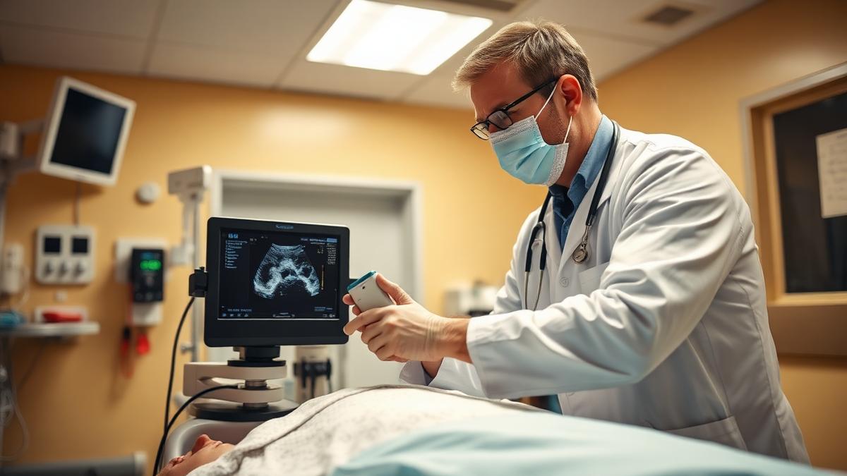
Instead of coordinating this complex transit, the clinical team wheels a device the size of a standard office photocopy machine directly to the patient's bedside. Plugged into a standard wall outlet, the machine scans the patient's head within minutes, transmitting high-resolution structural images directly to the hospital's picture archiving and communication system. This scenario is no longer theoretical. The convergence of Hyperfine's portable magnetic resonance imaging hardware and advanced software suites, such as those developed by Optive AI, is shifting the paradigm of neurocritical care diagnostics.
The Technical Breakthrough of Low-Field Imaging
Traditional magnetic resonance imaging systems operate at high magnetic field strengths, typically 1.5 Tesla or 3.0 Tesla. These systems require heavy structural reinforcement, extensive radiofrequency shielding, and liquid helium cooling systems to maintain superconductivity. Because of these constraints, conventional scanners are permanently anchored in specialized basement suites, far removed from the emergency department and the intensive care unit.
The alternative is low-field magnetic resonance imaging, which refers to diagnostic magnetic resonance scans performed at magnetic field strengths below 0.1 Tesla. Hyperfine's flagship system, the Swoop, utilizes a permanent magnet operating at 0.064 Tesla. This low field strength eliminates the need for expensive radiofrequency shielding and liquid helium cryogenics, allowing the device to be safely operated in the presence of nearby metallic objects and wheeled directly into active clinical spaces.
Operating at 0.064 Tesla presents a fundamental physical challenge: a severe reduction in signal strength. The signal-to-noise ratio is defined as the ratio of the desired signal power to the background noise power in an image. Because this ratio scales nonlinearly with magnetic field strength, a low-field scanner inherently collects far less raw physical data than its high-field counterparts. Under conventional reconstruction methods, this results in grainy, low-contrast images that lack the resolution necessary to make confident clinical decisions regarding subtle brain pathologies.
How Optive AI Bridges the Diagnostic Gap
To transform low-field scans into clinically actionable diagnostic tools, hardware advances must be paired with sophisticated computational methods. This is where specialized artificial intelligence platforms, including the reconstruction algorithms designed by Optive AI, come into play. By replacing traditional mathematical image reconstruction with neural networks trained on paired low-field and high-field datasets, software can infer missing structural details and filter out ambient electrical noise.
This computational process is called deep learning reconstruction, a method of generating clinical images by passing raw scanner data through trained deep neural networks to suppress artifacts and enhance structural boundaries. The AI does not invent or hallucinate anatomical structures; instead, it uses learned prior distributions of human brain anatomy to resolve the true physical boundaries obscured by noise.
Optive AI's software integration specifically addresses the types of noise found in clinical environments. An intensive care unit is filled with electromagnetic interference from ventilators, telemetry monitors, and infusion pumps. Traditional MRI scanners rely on a copper-shielded Faraday cage to block this external noise. Bedside scanners do not have this luxury. The AI algorithms must actively identify and subtract this environmental radiofrequency noise in real time, separating the patient's biological signals from the background hum of the ICU.
Operational and Financial Implications for Hospitals
The integration of bedside imaging and artificial intelligence redefines the economics of acute care. A conventional high-field suite requires an initial capital expenditure of several million dollars, supplemented by ongoing maintenance contracts and dedicated technical staff. For many community hospitals, the cost of installing and maintaining a second or third MRI scanner is prohibitive, leading to significant outpatient backlogs and delays in inpatient care.
Point-of-care imaging refers to medical diagnostic testing performed at or near the site of patient care, eliminating the need to transport the patient or specimens to a centralized laboratory or department. By adopting this approach for neuroimaging, hospitals can preserve their high-field scanners for complex, elective outpatient cases, such as tumor staging or functional cardiac imaging, which yield higher reimbursement rates and require extreme spatial resolution.
Using bedside units for routine follow-up scans, such as measuring ventricular size after shunt placement, frees up valuable high-field scanner time. This improves overall hospital throughput and reduces the length of stay in the intensive care unit. A patient waiting twelve hours for a routine head scan occupies an ICU bed that could otherwise host a new admission. Accelerating the imaging pipeline directly correlates with improved bed utilization metrics.
Furthermore, the staffing requirements for bedside scans are minimal. Because the magnetic field of a 0.064 Tesla scanner is highly localized, the strict safety perimeter required for high-field magnets is unnecessary. Intensive care nurses and resident physicians can remain in the room with the patient during the scan, eliminating the need for specialized MRI technicians to manage complex patient safety screenings.
Navigating Clinical Limits and the Path Forward
Despite these computational enhancements, low-field bedside imaging is not a universal replacement for high-field MRI. It is a complementary tool designed for specific, high-value clinical scenarios. High-field systems remain the gold standard for identifying subtle white matter lesions, characterizing complex skull base tumors, and conducting detailed vascular imaging.
The primary objective of bedside imaging combined with Optive AI is rapid, serial assessment. In conditions such as traumatic brain injury, ischemic stroke, and hydrocephalus, clinicians do not always need to see microscopic tissue details. They need to know if a midline shift has worsened, if a hemorrhage has expanded, or if ventricles are dilating. By providing these answers at the bedside within thirty minutes, the clinical team can make immediate decisions regarding surgical intervention or medical management, avoiding unnecessary interventions and reducing patient risk.
As software models continue to improve through exposure to larger, more diverse clinical datasets, the boundary of what can be resolved at low-field strengths will expand. The collaboration between flexible hardware developers like Hyperfine and software innovators like Optive AI demonstrates that the future of medical imaging lies not just in stronger magnets, but in smarter mathematics.
Key Signals
The adoption of bedside MRI systems demonstrates that computational power can effectively substitute for heavy physical infrastructure in medical diagnostics.
By offloading routine neurological monitoring to portable bedside scanners, health systems can optimize their high-field imaging assets for high-revenue outpatient procedures.
The ongoing validation of artificial intelligence reconstruction software by regulatory bodies establishes a precedent where software quality is as critical to clinical efficacy as hardware specifications.
As clinical proof points accumulate, bedside neuroimaging will transition from an innovative intensive care luxury to a standard operating capability in emergency departments and stroke centers worldwide.