An interventional cardiologist walks into the cath lab with a plan. Today, for most patients, that plan is a sketch. The angiogram shows where the narrowing is, but not how much calcium sits behind it, how long the diseased segment really runs, or what size the healthy vessel is on either side. So for complex cases the operator stops, threads an ultrasound catheter into the artery, measures, and decides. That pause is intravascular ultrasound, IVUS, and the evidence says it helps. It also costs time, contrast, a disposable catheter and a specialist who can read the images.
P4, presented as a late-breaking trial at TCT 2026 on November 1 and published the same day in NEJM, asked a simple question: what if most of that measuring happened before the patient arrived, from a coronary CT scan? The answer, in 1,105 randomized patients, was that the CCTA-guided strategy met noninferiority versus IVUS at 12 months, with shorter procedures and less contrast. Below we set out what the trial showed, where it sits on the evidence ladder, who gains and who is exposed, and what would have to be true for this to change everyday practice.
The question is no longer whether CT can find coronary disease. It is whether CT can decide how to treat it before anyone picks up a catheter.
In This Deep Dive
- Why this matters now
- What actually happened
- The obvious read versus the deeper signal
- Category map and taxonomy
- Evidence Ladder
- Regulation, payment and international divergence
- Capital flows and bottom-up economics
- The HealthTech Investor's Signal
- Implementation considerations
- Forward Intelligence
- The Bottom Line
Why this matters now
Coronary CT angiography has spent the last decade moving up the diagnostic pathway. In chest pain workups it has become a first-line test in many guidelines, and software that estimates fractional flow reserve from CT, known as FFRCT, has given it a physiological dimension that a plain anatomical picture lacks. What CT has not done, at scale, is reach into the procedure itself. The plan for a stent, which vessel, how long, what diameter, whether to modify calcium first, has stayed inside the cath lab, made by an operator looking at an angiogram and, in the best centers, an intravascular image.
Meanwhile, the case for intravascular imaging has strengthened. Over several years, randomized trials and meta-analyses have linked imaging-guided PCI to fewer adverse events than angiography alone, especially in complex lesions, and recent European and US guidance recommends intravascular imaging for complex lesions such as left main and long or calcified disease. Yet adoption remains uneven. Imaging adds workflow steps and requires training, and many labs use it for a minority of cases. That gap between what the evidence recommends and what labs actually do is exactly where a planning tool that works before the procedure could matter.
The timing is also commercial. Heartflow, which listed on Nasdaq under HTFL, reports use of its products at more than 1,800 institutions and in more than 750,000 patients. It has moved from a single diagnostic product, FFRCT, toward a portfolio that includes plaque analysis and procedural planning. P4 is the first large randomized test of whether that planning layer belongs in the treatment pathway, not just the diagnostic one.
Certainty: (A) Heartflow's reported footprint is company data; (B) our summary of the intravascular imaging evidence and guideline direction is general and not drawn from a single source; (B) the adoption gap is widely reported but varies by country and center.
What actually happened
Heartflow announced the results via GlobeNewswire on November 1 at 13:45 ET, and the American College of Cardiology published a summary the same day. The trial is registered as NCT05253677. It was investigator-initiated and supported by Heartflow.
The design
- Population: 1,105 patients with stable coronary artery disease and a clinical indication for revascularization, at least one epicardial lesion with a diameter stenosis of more than 70% and a fractional flow reserve of 0.80 or less, per ACC. Median age was 65, and nearly 22% were women.
- Sites: 23 centers in the United States, Europe and Japan.
- Randomization: PCI planned and guided by coronary CT angiography, or PCI guided by IVUS.
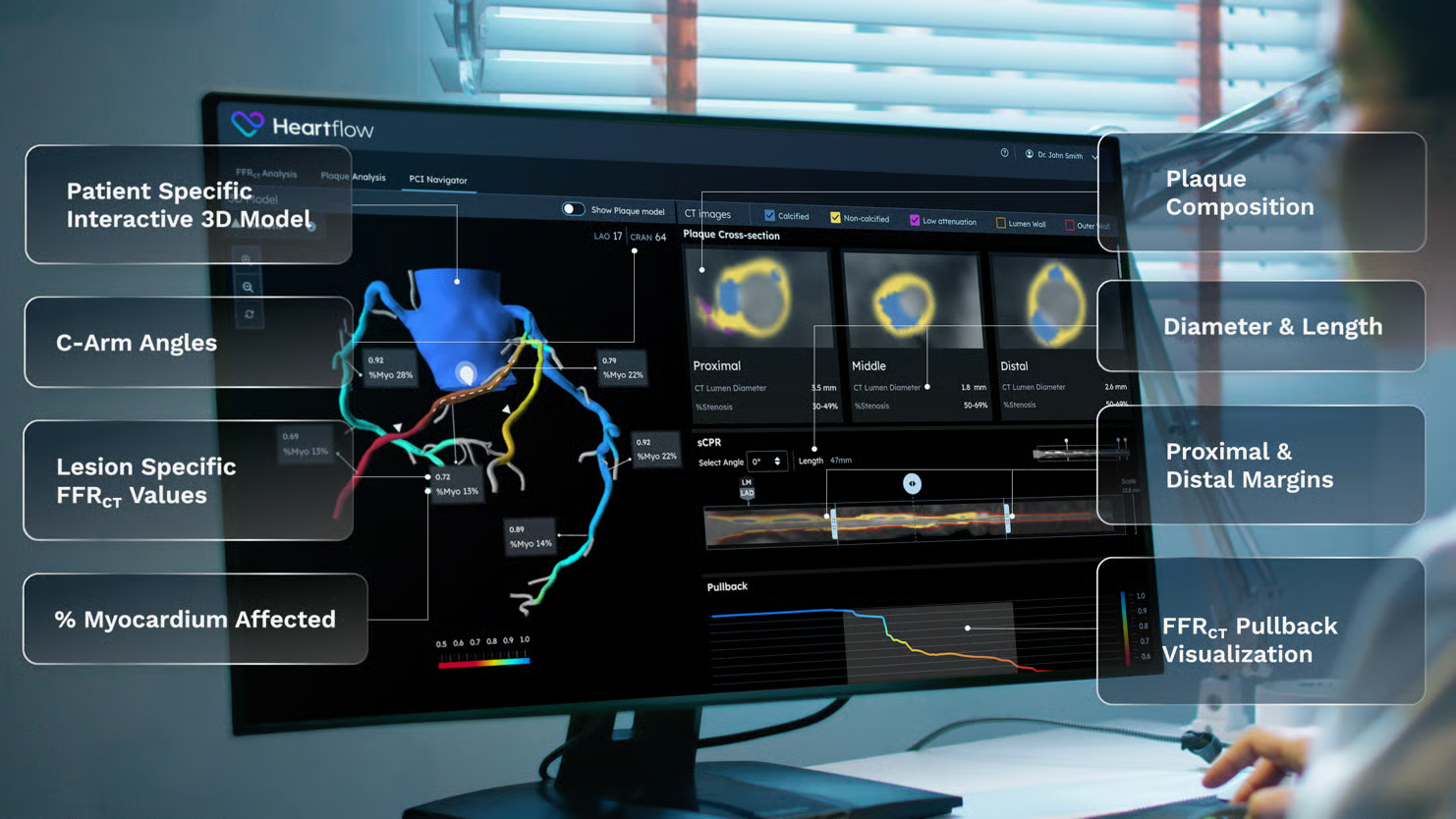
- CT tools: Heartflow FFRCT Analysis and Heartflow Planner, which the company describes as an earlier version of its PCI Navigator product.
- Primary endpoint: target-vessel failure at 12 months, tested for noninferiority.
The results
The primary endpoint is reported by ACC. Procedure time, contrast and calcium-modification measures below are attributed to Heartflow’s cited release.
- Target-vessel failure: 23 patients (4.1%) in the CT arm vs 25 patients (4.6%) in the IVUS arm. Noninferiority was met.
- Procedure time: median 46 vs 63 minutes, 17 minutes shorter, a 27% reduction.
- Contrast: median 150 vs 170 mL, about 12% less.
- Calcium modification: advanced calcium modification was used in 9.2% of CT-arm patients vs 3.8% in the IVUS arm.
- Complications: clinical procedural complications in 6 CT-arm patients vs 9 in the IVUS arm, per ACC.
What the investigators said
Principal investigator Carlos Collet, MD, PhD, of the Cardiovascular Research Foundation, framed the comparison carefully: "Intravascular ultrasound is an established benchmark, but performing assessments inside the cath lab adds procedural time and requires expertise." He continued: "P4 shows that non-invasive CT-guided PCI planning achieves comparable clinical outcomes with shorter procedure times and less contrast, enabling interventionalists to anticipate challenges, tailor their strategy, and enter the cath lab better prepared."
The ACC summary quotes the investigators' published conclusion: "The findings suggest that key PCI-planning decisions can be shifted from the cardiac catheterization laboratory to preprocedural CCTA, thereby extending the role of CCTA beyond diagnosis." They add that upstream planning creates "the opportunity to enhance shared decision-making before the invasive procedure."
Heartflow Chief Medical Officer Campbell Rogers, MD, drew the structural heart comparison: "CT has already become routine for planning structural procedures like TAVR." That is the right analogy to test, and we come back to it below.
The companion data
Heartflow also presented NAVIGATE-PCI, a 203-patient registry led by Ziad A. Ali, MD, DPhil, of the DeMatteis Cardiovascular Institute at St. Francis Hospital. Physicians first planned with the angiogram alone, then reviewed PCI Navigator. Plans changed in about 70% of patients, most often stent length (41%) and lesion preparation (12%). A third dataset, BRAVEHEART, covering 96 firefighters with at least 10 years of smoke exposure, is scheduled at TCT on November 3 and concerns prevention rather than PCI.
Certainty: (A) design, endpoint and results as reported by Heartflow and ACC; (A) quotes as published; (B) NAVIGATE-PCI is a single-arm registry and its plan-change figure measures decisions, not outcomes.
The obvious read versus the deeper signal
The obvious read: The CCTA-guided strategy met noninferiority versus IVUS, with a company-reported 17-minute difference in median procedure time. Heartflow has a new product with randomized support, and interventional cardiologists have a reason to order a CT before elective PCI. All of that is true, and it is good news for a company that has spent years building the evidence base for FFRCT.
The deeper signal: look at where the decision is made. In the IVUS arm, the plan is made inside the procedure, by the operator, using a disposable catheter. In the CT arm, the plan is made before the procedure, by software and a reader, using a scan that may already exist from the diagnostic workup. That is not a small change in tooling. It moves the decision point upstream, out of the most expensive room in the hospital.
Three implications follow. First, the work changes hands. Someone has to read the CT plan, check it and own it. That could be the interventional cardiologist the night before, an imaging cardiologist, or a dedicated planning service. Structural heart programs went through exactly this shift with TAVR, where CT planning became routine and a CT reading role grew around it. Second, the cath lab becomes more predictable. If calcium is known in advance, the operator can bring the right tools, and the higher use of advanced calcium modification in the CT arm (9.2% vs 3.8%) suggests that is already happening. Predictable cases are easier to schedule and staff. Third, the evidence bar for the incumbents rises. IVUS and optical coherence tomography (OCT) vendors now need to show what they add when a good CT plan already exists, not just what they add over angiography alone.
There is a quieter fourth point. The NAVIGATE-PCI figure, plans changed in about 70% of patients, is striking but should be read with care. A tool that changes plans often is only valuable if the changed plans are better. P4 tested outcomes against IVUS, not against angiography with or without the planner. The plan-change number shows the tool is influential; P4 shows that a CT-guided strategy does not do worse than the imaging benchmark. Those are different claims, and both matter.
Certainty: (A) the arm structure and calcium modification rates; (B) the TAVR analogy and workforce implications are our interpretation; (C) forward-looking view on scheduling and staffing.