In acute cardiovascular medicine, the clinical adage "time is muscle" has driven massive health system investments over the past two decades. Interventional cardiology networks and emergency departments across the United States have successfully optimized in-hospital "door-to-balloon" times, routinely achieving percutaneous coronary intervention (PCI) within 60 minutes of patient arrival.
Yet, despite these hospital-based efficiencies, overall mortality and long-term heart failure rates following acute myocardial infarction (MI) remain stubbornly elevated. The reason is a severe systemic failure in the pre-hospital window: the average "symptom-to-door" time has remained stalled at 2 to 4 hours for over thirty years. Patients experiencing ambiguous chest discomfort, atypical symptoms, or mild angina frequently hesitate, wait for symptoms to subside, or drive themselves to urgent care centers rather than activating emergency medical services (EMS).
In This Deep Dive:
- Why this matters now: The pre-hospital time gap in acute coronary syndromes.
- What actually happened: FDA Breakthrough Device Designation awarded within 30 days.
- The obvious read versus the deeper signal: Consumer ECG wearables vs synthesized 12-lead vectorcardiography.
- Competitive taxonomy & clinical maturity: Ambulatory and emergency cardiac diagnostics.
- The Evidence Ladder: 3D dipole physics to at-home emergency medical activation.
- 📈 The HealthTech Investor's Signal: Remote diagnostic RPM billing, health system risk sharing, and commercial TAM.
- Counter-thesis: False positive EMS overload, patient placement artifacts, and telemetry liability.
- Forward intelligence: 4 observable test indicators for the upcoming 12 to 24 months.
- The bottom line for health system leaders, interventional cardiologists, and MedTech investors.
Why this matters now
Every year, more than 805,000 Americans suffer a heart attack, with coronary heart disease remaining the number one cause of death globally. Over 20 million high-risk Americans live with diagnosed coronary artery disease, prior stents, or previous coronary artery bypass graft (CABG) surgery, living under continuous anxiety regarding potential ischemic recurrence.
Consumer single-lead and six-lead ECG wearables (such as Apple Watch or KardiaMobile) have achieved widespread adoption for detecting rhythm disturbances like atrial fibrillation. However, they are fundamentally incapable of diagnosing acute myocardial infarction because ST-segment elevation and acute ischemic vector shifts require a standardized 12-lead spatial projection across anatomical leads (I, II, III, aVR, aVL, aVF, and V1-V6). By securing FDA Breakthrough Device Designation for an at-home system that synthesizes standard 12-lead ECGs from a cable-free 3D vector sensor, HeartBeam directly targets this multi-hour diagnostic blind spot.
What actually happened
The FDA granted Breakthrough Device Designation to HeartBeam's 3D ECG at-home heart attack assessment system within 30 days of submission, an exceptionally rapid turnaround reflecting the critical unmet need in pre-hospital coronary triage.
The designation was supported by clinical evidence presented at the American Heart Association (AHA) Scientific Sessions and data from the 134-patient ALIGN-ACS pilot trial. The study demonstrated that HeartBeam's cable-free, pocket-sized device captures cardiac electrical signals in three non-coplanar orthogonal dimensions (X, Y, Z vectors) and uses proprietary mathematical synthesis algorithms to reconstruct a standard 12-lead ECG with high anatomical concordance to baseline hospital-grade 12-lead tracings.
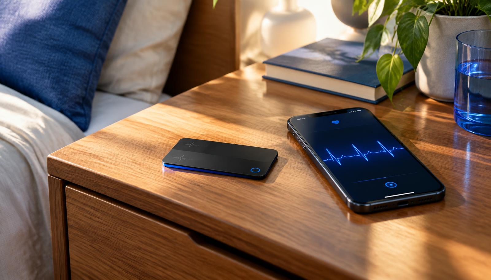
When a patient experiences symptoms, they place the credit-card-sized device against their chest for 30 seconds. The synthesized 12-lead data and automated AI ischemia detection algorithms immediately transmit to an on-call clinical reading center, enabling rapid emergency medical dispatch or reassuring non-ischemic resolution.
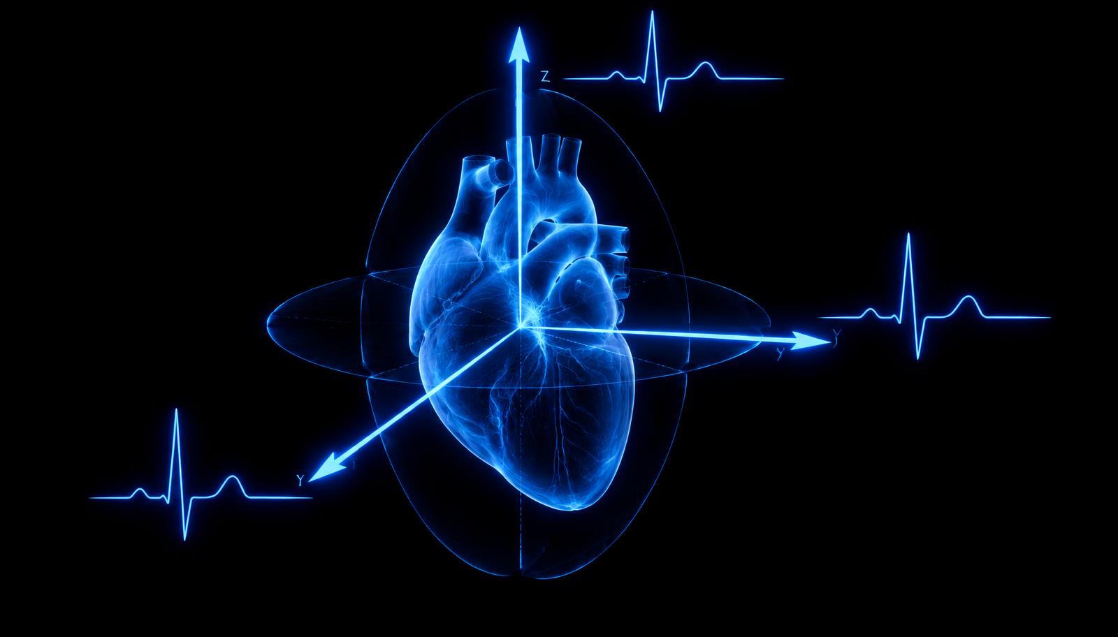
Vectorcardiography captures the heart's electrical dipole along three orthogonal axes. Image: The HealthTech Signal
The obvious read versus the deeper signal
The surface interpretation is that HeartBeam is releasing a high-end cardiovascular wearable to compete in the remote patient monitoring market. The deeper architectural signal is the transformation of vectorcardiography (VCG) from an obsolete academic technique into an AI-powered diagnostic engine.
Historically, capturing a 12-lead ECG required applying ten physical adhesive electrodes across specific anatomical landmarks, connected by a tangle of cables to a rolling hospital cart. This physical constraint locked 12-lead diagnostics inside clinical facilities. Vectorcardiography originally proved that all surface electrical activity originates from a single equivalent moving cardiac dipole that can be fully captured with three orthogonal axes. By combining 3D dipole physical sensing with deep-learning reconstruction algorithms, HeartBeam eliminates the physical cable harness while retaining the diagnostic fidelity of standard 12-lead ST-segment analysis. This moves hospital-grade triage directly to the patient's bedside.
🔒 HEALTHTECH SIGNAL PLUS · EXCLUSIVE SUBSCRIBER INTELLIGENCE