A sixty-seven-year-old patient sits in a primary care examination room, describing a subtle, occasional shortness of breath when walking uphill. In previous years, this presentation would prompt a referral to a cardiologist, initiating a waiting period of several weeks or months for a diagnostic transthoracic echocardiogram. Today, the primary care physician reaches for a handheld ultrasound probe connected to a tablet. Guided by real-time software prompts, the clinician obtains a diagnostic-quality view of the heart within two minutes. The integrated software immediately calculates a left ventricular ejection fraction of forty-two percent, indicating early-stage heart failure. This scenario is no longer an experimental pilot project; it is the practical clinical reality of cardiac diagnostics in 2026.
The Evolution of ECG Analysis from Detection to Prediction
For decades, computerized electrocardiogram analysis was limited to basic rhythm identification, such as flagging atrial fibrillation or acute myocardial infarction. Today, the capabilities of the resting twelve-lead ECG have expanded significantly. Opportunistic screening is the practice of utilizing routinely performed clinical tests to detect complex, asymptomatic structural heart diseases before clinical symptoms fully manifest.
Deep learning models trained on millions of paired ECG and echocardiogram datasets can now identify subtle electrical signatures associated with structural abnormalities. These include left ventricular dysfunction, silent hypertrophic cardiomyopathy, and moderate-to-severe aortic stenosis. In 2026, these algorithms run silently in the background of health system networks. When a clinician orders a routine ECG for a pre-operative evaluation or a wellness visit, the AI engine analyzes the waveform and inserts a risk score directly into the electronic health record.
This shift addresses a major clinical challenge. Many patients with structural heart disease remain asymptomatic until their condition reaches an advanced stage. By converting a low-cost, widely available test into a highly sensitive screening tool, health systems can identify high-risk patients earlier. This enables timely pharmacological intervention, which can slow disease progression and prevent costly hospitalizations.
Democratising Echocardiography at the Point of Care
Formal echocardiography remains a major diagnostic bottleneck in modern medicine. The shortage of credentialed cardiac sonographers has created severe backlogs, forcing patients to wait long periods for essential diagnostic imaging. While point-of-care ultrasound devices have been available for years, their utility was historically limited by the high level of technical skill required to acquire and interpret cardiac images.
To bridge this gap, modern diagnostic software utilizes advanced computer vision. Automatic view guidance refers to the real-time software assistance that instructs a non-specialist user on how to manipulate an ultrasound transducer to capture standardized diagnostic views.
The software provides on-screen prompts, directing the clinician to tilt, slide, or rotate the probe. Once the system detects an optimal apical four-chamber or parasternal long-axis view, it automatically records the loop and calculates key metrics, such as ejection fraction and stroke volume. For hospital administrators, this democratization of cardiac imaging improves patient throughput. By enabling emergency physicians, hospitalists, and primary care providers to perform reliable basic evaluations, specialized sonographers can dedicate their time to complex diagnostic studies, optimizing the overall efficiency of the imaging department.
Multimodal Fusion and Clinical Decision Support
While isolated algorithms for ECGs and ultrasounds are valuable, clinical cardiology requires a holistic view of the patient. The most significant architectural advancement in 2026 is the transition toward multimodal diagnostic models. Multimodal clinical fusion is the computational integration of disparate data streams, including continuous waveform data, medical imaging, laboratory values, and unstructured historical text, to generate a unified clinical assessment.
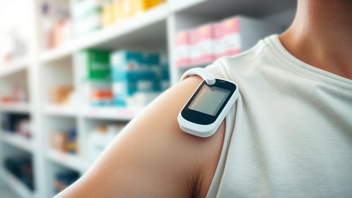
Instead of reviewing an isolated diagnostic report, clinicians now interact with systems that synthesize the patient's entire cardiac history. For example, a modern platform can combine a patient's historical blood pressure trends, their recent serum biomarker levels, and continuous physiological data from a wearable patch. The algorithm analyzes these data points collectively to predict the likelihood of a major adverse cardiac event or a heart failure exacerbation within a specific timeframe.
For technology founders and developers, this shift represents a move away from single-task utility toward comprehensive clinical workflow integration. The value of a diagnostic tool is increasingly measured by how seamlessly it integrates into existing workflows and whether it reduces the cognitive burden on clinicians by presenting consolidated, actionable insights rather than disparate data points.
Regulatory, Economic, and Workflow Integrations
The widespread adoption of AI in cardiac diagnostics has been accelerated by clear regulatory guidelines and established reimbursement pathways. The United States Food and Drug Administration has refined its oversight of software as a medical device, establishing predictable evaluation frameworks for algorithms that adapt to new clinical data.
Equally important is the progress in reimbursement. The American Medical Association has introduced Category I Current Procedural Terminology codes for AI-enabled analysis of physiological data. These codes allow clinical practices to bill for the algorithmic interpretation of ECG waveforms and automated echocardiographic assessments. This structural financial support has transformed cardiac AI from an administrative expense into a viable revenue-generating asset for healthcare practices.
Despite these advancements, operational challenges persist. Clinicians are hesitant to rely on algorithms that do not provide clear explanations for their outputs. In response, diagnostic platforms are incorporating explainable interface designs, such as highlighting the specific segments of an ECG waveform or the precise myocardial walls in an ultrasound loop that influenced the algorithm's calculation. Ultimately, these tools are designed to function as decision support systems, keeping the licensed clinician as the final decision-maker to manage clinical liability and ensure patient safety.
Key Signals
The decentralization of cardiac diagnostics from specialized imaging labs to the clinical point of care is significantly reducing diagnostic wait times and enabling earlier therapeutic intervention.
The establishment of dedicated medical billing codes for algorithmic analysis has provided the financial foundation necessary for wide-scale clinical adoption across diverse healthcare settings.
Successful healthcare technology developers are prioritizing the seamless integration of multimodal data into existing electronic health records to minimize clinician administrative burden and prevent alert fatigue.
The clinical utility of cardiac artificial intelligence is increasingly defined by its ability to predict acute cardiovascular events rather than merely classifying existing chronic conditions.