In a quiet primary care clinic in suburban Ohio, a family physician reviews a standard twelve lead electrocardiogram printout for a sixty-two year old patient presenting for a routine annual physical. To the physician's eye, the waveforms appear unremarkable. The rhythm is sinus, the intervals are within normal limits, and there are no obvious ST-segment deviations or pathological Q waves. Under traditional clinical guidelines, this tracing would be filed away as normal, and the patient would be sent home with a clean bill of cardiovascular health.
However, when this same tracing is processed by an artificial intelligence algorithm trained on millions of historical electrocardiograms, the system flags the patient as high risk for having a reduced left ventricular ejection fraction, which is defined as the percentage of blood pumped out of the left ventricle with each contraction. A subsequent echocardiogram confirms the algorithm's prediction, revealing a silent, early-stage cardiomyopathy that can now be managed with beta-blockers and sodium-glucose cotransporter-2 inhibitors long before the patient develops clinical symptoms of heart failure.
This scenario represents the practical promise of AI-enabled electrocardiography. By extracting imperceptible patterns from raw electrical signals, machine learning algorithms are converting a ubiquitous, inexpensive, and century-old diagnostic tool into a highly sensitive screening device for structural and functional heart diseases.
The Diagnostic Gap in Standard Electrocardiography
For decades, the electrocardiogram (ECG) has served as the frontline diagnostic tool for acute cardiac events, such as myocardial infarction, and overt rhythm disturbances. Yet, its utility in detecting structural heart disease has been severely limited. A standard twelve lead ECG offers a brief ten second snapshot of the heart's electrical activity. While human cardiologists are highly skilled at identifying clear abnormalities like bundle branch blocks or acute ischemia, they cannot reliably detect the minute, diffuse changes in voltage and timing that indicate early ventricular remodeling or valvular dysfunction.
This limitation leaves millions of patients undiagnosed during the early phases of progressive cardiac conditions. For instance, asymptomatic left ventricular dysfunction is a condition where the heart's left ventricle is weakened but the patient does not yet exhibit overt symptoms of heart failure. It affects up to seven percent of the global population over the age of sixty-five. Because patients remain asymptomatic, the condition is typically caught only after irreversible damage has occurred or when a patient presents to an emergency department with acute shortness of breath.
A similar diagnostic challenge exists for hypertrophic cardiomyopathy, which is defined as a genetic disease in which the heart muscle becomes abnormally thick, making it harder for the heart to pump blood. Traditional ECG criteria for hypertrophy have poor sensitivity, missing a substantial portion of affected individuals who remain at risk for sudden cardiac death. The inability of human clinicians to extract these subtle structural indicators from standard ECG waveforms represents a major diagnostic gap in preventive cardiology.
The Mechanics of AI-Enabled ECG Analysis
Artificial intelligence bridges this gap by shifting the paradigm from manual rule-based interpretation to pattern recognition across thousands of data points. Rather than relying on simple measurements like the PR interval or QRS duration, these models analyze the raw digitized voltage-time data across all twelve leads simultaneously.
At the core of this technology are deep learning networks, which are defined as a subset of machine learning based on artificial neural networks with multiple layers that can learn complex representations from raw input data. Specifically, convolutional neural networks are trained on pairs of ECG tracings and gold-standard imaging data, such as echocardiograms or cardiac magnetic resonance imaging. During the training process, the network identifies subtle, spatial-temporal patterns across leads that correlate with structural changes, such as myocardial fibrosis, chamber enlargement, or localized wall motion abnormalities.
These algorithmic models can detect signature patterns of heart disease that are invisible to human experts. For example, a model can identify the specific electrical signature of a heart that is struggling to pump efficiently even when the overall rhythm and visible waveform structure appear completely normal to a board-certified cardiologist. This capability transforms the standard ECG into a low-cost, non-invasive surrogate for expensive and resource-intensive imaging studies.
Clinical Validation and Real-World Evidence
The clinical utility of AI-ECG is no longer merely theoretical. Extensive academic validation has demonstrated that these algorithms can identify patients with reduced ejection fraction with high accuracy. In landmark studies, models trained on large clinical datasets achieved an area under the receiver operating characteristic curve of over zero point nine zero, demonstrating diagnostic performance comparable to or exceeding many widely accepted screening tests, such as mammography for breast cancer or prostate-specific antigen testing for prostate cancer.
Crucially, real-world prospective validation has confirmed that these algorithmic insights translate to better patient care. In the pragmatically designed EAGLE trial, which was a cluster-randomized controlled trial conducted in primary care practices, clinics that had access to an AI-ECG screening tool identified significantly more patients with low ejection fraction than clinics utilizing standard care. The intervention did not lead to an unmanageable surge in diagnostic downstream testing, suggesting that the tool successfully guided clinicians toward the patients who truly needed echocardiographic evaluation.
Furthermore, the technology has shown remarkable efficacy in detecting silent atrial fibrillation, which is defined as episodes of irregular heart rhythm that occur without recognizable symptoms. By analyzing a single ECG recorded during normal sinus rhythm, AI models can identify patients with a high probability of experiencing paroxysmal atrial fibrillation episodes at other times. This allows clinicians to initiate anticoagulation therapy or closer monitoring before a patient experiences a devastating embolic stroke.
Operational Integration and Workflow Design
For healthtech operators and clinical leaders, the primary challenge of AI-ECG lies not in the performance of the algorithm itself, but in its integration into the clinical workflow. If an AI tool requires clinicians to log into a separate portal, copy and paste data, or manually upload ECG files, adoption will remain minimal.
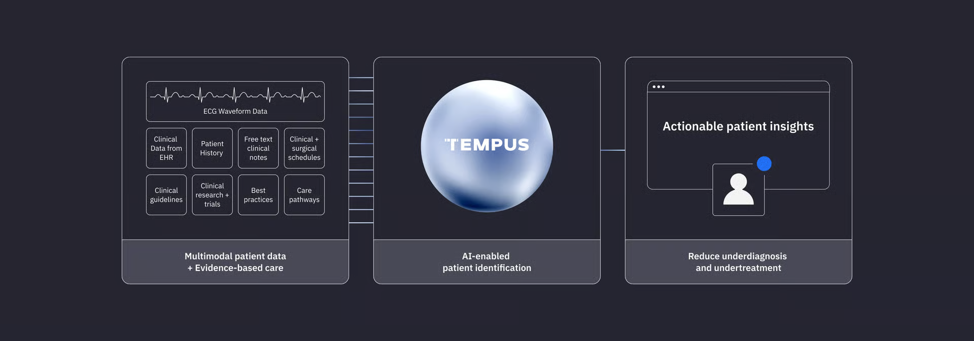
Successful implementation requires the algorithm to run silently in the background of existing electronic health record (EHR) systems. When an ECG is performed in a primary care clinic or urgent care center, the raw digital data should automatically flow to the cloud-based or on-premise AI engine. The resulting risk score should then populate directly within the clinician's native viewing window, ideally alongside a clear, actionable recommendation, such as ordering a point-of-care echocardiogram or referring the patient to a cardiologist.
Additionally, health systems must carefully calibrate the alert thresholds of these models. Setting the threshold too low will maximize sensitivity but generate a high rate of false positives, which can overwhelm local cardiology clinics and lead to unnecessary, costly imaging studies. Conversely, setting the threshold too high will miss critical cases of early-stage heart disease. Clinical operations leaders must establish clear pathways for managing positive results, ensuring that primary care physicians have rapid access to secondary testing and specialist consultation to manage the identified risk appropriately.
Key Signals
The transformation of the standard electrocardiogram into an advanced screening tool represents a fundamental shift in cardiovascular care from reactive treatment to proactive, early-stage intervention.
For clinical systems, successful deployment of AI-ECG hinges on designing silent, integrated workflows within the electronic health record to prevent cognitive fatigue among front-line clinicians.
Healthtech innovators must focus on validating these algorithms across diverse patient populations to ensure that diagnostic accuracy remains consistent across varying age groups, sexes, and ethnicities.