At a community health clinic in eastern Iowa, a medical assistant guides a 54 year old patient with type 2 diabetes toward a specialized fundus camera. The patient has not seen an ophthalmologist in four years. The assistant captures two images of each retina, uploads them to an automated system, and within sixty seconds, the clinic workstation displays a clear result: referable diabetic retinopathy detected. No specialist has looked at the images, yet the diagnosis carries the clinical weight of a board-certified eye surgeon.
This scenario represents the operational reality of clinical artificial intelligence in 2026. The technology has matured past the experimental phase and has integrated into the structural fabric of daily clinical workflows. For healthtech operators, clinicians, and health system executives, the question is no longer whether to adopt diagnostic AI, but how to deploy it to maximize clinical throughput, secure reimbursement, and improve patient safety.
To evaluate the leading tools in this space, we must look beyond venture capital funding or marketing claims. The critical metrics are clinical validation, integration into existing electronic health records, and established reimbursement pathways. The following diagnostic AI tools represent the highest standard of clinical utility and operational maturity in 2026.
1. LumineticsCore
LumineticsCore, formerly known as IDx-DR, is an autonomous diagnostic system designed to detect diabetic retinopathy in primary care settings. The system analyzes high-resolution images of the retina taken with a non-mydriatic fundus camera. By enabling primary care clinics to test patients during routine checkups, the technology bypasses the traditional bottleneck of specialist referrals.
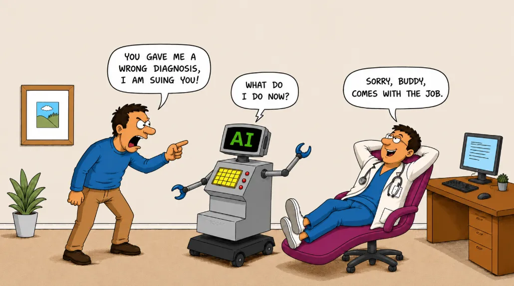
To understand how this system operates, it is helpful to define its core architecture. Autonomous AI is defined as software that analyzes clinical data and generates a diagnostic decision without human physician intervention. Under this model, the software assumes the legal and clinical responsibility for the diagnostic output, which distinguishes it from traditional decision-support systems.
The diagnostic accuracy of the platform is backed by a pivotal clinical trial involving several hundred patients, where it demonstrated high sensitivity and specificity in detecting diabetic retinopathy. The system integrates directly into popular electronic health records, allowing the automated report to populate the patient chart automatically.
Why it matters
For primary care clinics, this technology transforms a cost center or a lost referral into an immediate revenue generator through the CPT code 92229. It increases the compliance rate for diabetic eye exams, which is a key quality metric for health systems participating in value-based care contracts. More importantly, it catches sight-threatening pathology in patients who would otherwise never see an eye specialist.
2. Viz LVO
Viz LVO is an automated triage system that analyzes computed tomography angiograms of the brain to identify suspected large vessel occlusions. These occlusions are the most severe type of ischemic stroke and require rapid intervention to prevent permanent disability. The software runs in the background of the hospital imaging network, scanning every eligible scan as soon as it is completed.
To clarify its role in the emergency department, we must understand its classification. Computer-aided triage is defined as software that analyzes medical images to identify suspected critical findings and immediately alerts the appropriate specialist. Rather than forcing the specialist to wait for the general radiologist to read the scan, the system alerts the neurointerventional team via a secure mobile application.
The platform has been shown to reduce the time from patient arrival to mechanical thrombectomy. In stroke care, where every minute of delay results in the loss of millions of cortical neurons, this acceleration in care delivery changes clinical outcomes.
Why it matters
For hospital networks, Viz LVO acts as a coordination hub that connects community hospitals with comprehensive stroke centers. The automated alert system ensures that transfer decisions are made in minutes rather than hours. This optimization of the referral pathway increases the volume of appropriate interventional procedures at tertiary centers while ensuring community patients receive timely care.
3. Cleerly Coronary Clinical Suite
The Cleerly Coronary Clinical Suite is an AI-powered platform that analyzes coronary computed tomography angiography scans to identify and characterize coronary artery disease. Traditional interpretations of these scans focus on the degree of vessel narrowing, which can be a poor predictor of actual cardiac events. Cleerly shifts the focus to the pathology of the vessel wall itself.
To comprehend the diagnostic value of this approach, we must examine the specific pathology it measures. Vulnerable plaque is defined as a lipid-rich coronary lesion that is highly susceptible to rupture and subsequent myocardial infarction. By identifying and quantifying this specific tissue type, the software allows clinicians to identify patients at high risk of a heart attack long before they present with classic symptoms.
The system provides a three-dimensional reconstruction of the coronary arteries, color-coding the vessel walls based on the type of plaque present. This precise quantification allows cardiologists to track the progression or regression of disease over time in response to medical therapies.
Why it matters
This technology changes the clinical management of coronary artery disease from reactive revascularization to proactive prevention. It provides clinicians with objective, quantitative data to justify aggressive lipid-lowering therapies or, conversely, to avoid unnecessary invasive cardiac catheterizations in stable patients. This capability aligns perfectly with the goals of risk-bearing healthcare organizations.
4. HeartFlow FFRCT Analysis
HeartFlow FFRCT Analysis is a non-invasive diagnostic tool that assists clinicians in evaluating patients with suspected coronary artery disease. The software utilizes deep learning algorithms and computational fluid dynamics to calculate pressure drops across coronary stenoses from standard coronary computed tomography angiography images.
This process relies on a key physiological metric. Fractional flow reserve is defined as the ratio of maximum blood flow in a stenotic artery to the maximum flow in a normal artery. Historically, measuring this ratio required an invasive cardiac catheterization procedure, where a pressure-wire was threaded directly into the coronary artery. HeartFlow calculates this metric non-invasively, producing a color-coded three-dimensional model of the coronary tree.
The clinical evidence supporting this technology is extensive, demonstrating that the use of non-invasive fractional flow reserve calculations significantly reduces the rate of negative invasive angiograms. It provides both structural and functional information from a single non-invasive scan.
Why it matters
By reducing the number of diagnostic catheterizations that show no obstructive disease, HeartFlow helps hospital systems optimize the use of their cardiac catheterization laboratories for therapeutic interventions rather than diagnostic procedures. This improves laboratory throughput, lowers overall procedural risks for patients, and reduces the total cost of care for payers.
5. Koios DS
Koios DS is a decision-support software platform designed to assist radiologists in interpreting ultrasound images of the breast and thyroid. Ultrasound interpretation is notoriously subjective, leading to high rates of unnecessary biopsies for benign lesions. The software uses deep learning models trained on millions of confirmed clinical images to analyze the characteristics of detected nodules.
This tool addresses a major challenge in diagnostic imaging. Inter-observer variability is defined as the difference in diagnostic assessments made by two or more clinicians evaluating the same clinical data. By providing an objective, secondary assessment that aligns with standard reporting systems, the software helps standardize clinical decision-making across an entire radiology department.
The system integrates directly into the picture archiving and communication systems used by radiologists. When a clinician highlights a lesion, the software analyzes its shape, margins, and echogenicity, generating a likelihood of malignancy within seconds.
Why it matters
The primary clinical benefit of this system is the reduction of unnecessary biopsies for benign lesions, which are a major source of patient anxiety and healthcare expenditure. For radiology practices, the software serves as a valuable second opinion that improves diagnostic confidence, accelerates reading times, and mitigates the risk of diagnostic errors.
Key Signals
The adoption of autonomous diagnostic systems in primary care clinics is shifting the point of care, allowing early disease detection and treatment to occur before patients require expensive specialist interventions.
The integration of automated triage tools into acute hospital networks is standardizing emergency response times and ensuring that patients with critical conditions are directed to appropriate interventions without delay.
The transition from subjective visual assessments to objective, quantitative plaque and tissue analysis is enabling personalized, preventative treatment strategies that can be tracked over time to measure clinical efficacy.