This account is based on reporting by The Independent and the Manchester Evening News. The HealthTech Signal has not independently interviewed Ms. Cook.
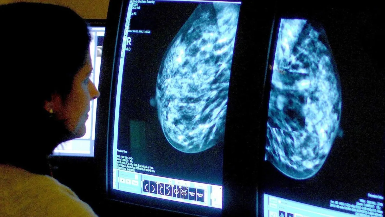
Yvonne Cook went in for a routine mammogram expecting the same result she had gotten before: normal, see you in three years. Instead, an AI system reviewing her scan alongside the radiologists flagged something the human readers had not caught, a tumor so small it had eluded the standard reading entirely. The Independent reported that the cancer was an aggressive form, one that Cook has said she believes would not have been caught until her next scheduled screening, potentially three years later, by which point it likely would have grown and spread.
Cook has publicly credited the AI with sparing her from what she called arduous cancer treatment, framing the early catch as the difference between a manageable intervention now and a much harder fight later. The Manchester Evening News quoted her saying she felt lucky the technology caught something so small at exactly the right time.
How AI actually reads a mammogram
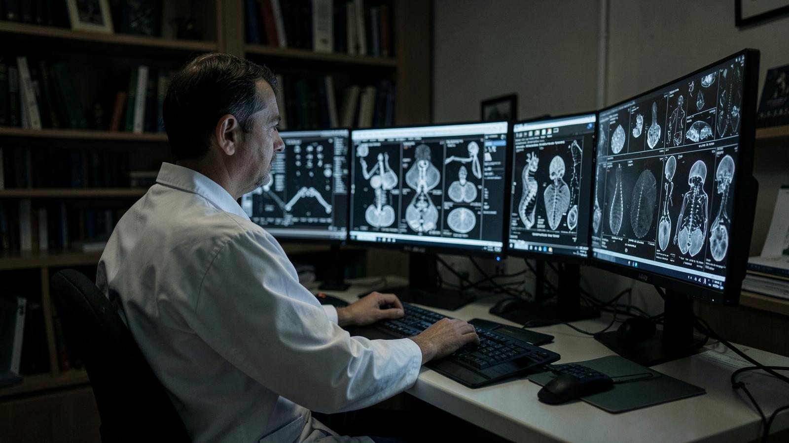
The tools now deployed across breast screening programs, including the one involved in Cook's case, work as a second reader alongside human radiologists, not a replacement for them. A radiologist reviews the images and renders a judgment. The AI system independently analyzes the same images using a model trained on millions of prior mammograms paired with confirmed outcomes, looking for subtle patterns of tissue density, calcification and architectural distortion that correlate with malignancy.
The clinical value shows up specifically in cases like Cook's: small, early-stage lesions in dense breast tissue, which is notoriously difficult for the human eye to read because dense tissue and tumors can appear similarly white on a mammogram image. This is precisely the population where independent research, including studies referenced in NBC News' reporting on AI mammography, has found AI systems can increase detection meaningfully, with some estimates suggesting AI-assisted reading increases breast cancer detection rates by more than 10 percent compared with human reading alone.
Why this is not simply "AI beats doctors"
I want to resist the framing that would turn this into a story about human radiologists failing. Mammography is genuinely one of the hardest pattern recognition tasks in medicine. Radiologists read enormous volumes of scans, most of which are normal, looking for signals that can be a few millimeters across and easily mimicked by ordinary tissue. Missed cancers on mammograms are a known, studied phenomenon that predates AI by decades, and interval cancers, tumors diagnosed between scheduled screenings, are exactly the category AI is proving useful against.
What AI changes is not the ceiling of what is detectable, it changes the consistency. A radiologist's performance can vary with fatigue, caseload, and the sheer volume of scans reviewed in a shift. An AI model applies the same pattern recognition to every image, every time, without fatigue. Used as a second reader, it functions less like a smarter doctor and more like a tool that never has an off day.
The part that deserves more scrutiny
The same NBC News reporting flagged the concern that responsible coverage of this technology has to include: overdiagnosis. An AI system tuned to catch every possible early signal will inevitably flag some abnormalities that would never have become clinically significant, findings that lead to biopsies, anxiety, and sometimes treatment for something that posed little real threat. The tradeoff between catching genuinely dangerous cancers early and over-treating harmless ones is not new to AI, it is the central tension of cancer screening generally, but AI's higher sensitivity can sharpen it in either direction depending on how the tool is calibrated.
There is also, as NBC's reporting noted, a meaningful gap between promising retrospective study results and prospective, real-world evidence that AI-assisted screening actually reduces mortality at a population level, rather than simply detecting more findings. Detection is not the same outcome as saved lives, and the trials capable of proving the latter take years to mature.
The systemic tension
Cook's case is a powerful individual story, and it is also, structurally, a story about screening infrastructure. National screening programs, including the NHS breast screening program referenced in this reporting, operate on fixed intervals, typically every three years in the UK, because screening more often than that has historically not been shown to justify the cost and radiation exposure involved for the average patient. AI does not change that interval. What it changes is the odds that a cancer present at the scheduled scan gets caught the first time, rather than waiting for the next one.
That is a real gain, but it is also unevenly distributed. AI-assisted mammography requires the imaging infrastructure, the software licensing, and the radiologist workflow integration to deploy at scale, and health systems are adopting it at different speeds. A patient screened at a center that has integrated AI reading gets a meaningfully different chance of early detection than a patient screened at a center that has not, even though both may be following the same national screening guideline.
The takeaway
Yvonne Cook's tumor was there on the image the whole time. What changed was not the biology, it was who, or what, was looking closely enough to see it. That is the honest, undramatic truth about AI in cancer screening: it is not magic, and it does not replace the radiologist. It is a second, tireless set of eyes on an image where a few millimeters can be the difference between an early intervention and a much harder diagnosis. The work now is making sure that second set of eyes is looking at every scan, not just the ones at hospitals that could afford to install it first.
Key Signals
AI-assisted mammography reading is showing measurable gains against a specific, well-documented failure mode: small, early-stage cancers hidden in dense breast tissue that human readers can miss. The technology functions as a tireless second reader rather than a replacement, and its main value is consistency rather than superior judgment. Overdiagnosis remains a real and unresolved tradeoff, since higher sensitivity will also flag findings that would never have become clinically significant. Access to this technology is currently uneven across health systems, so a patient's odds of early detection still depend partly on which center performs the scan.