Listen instead
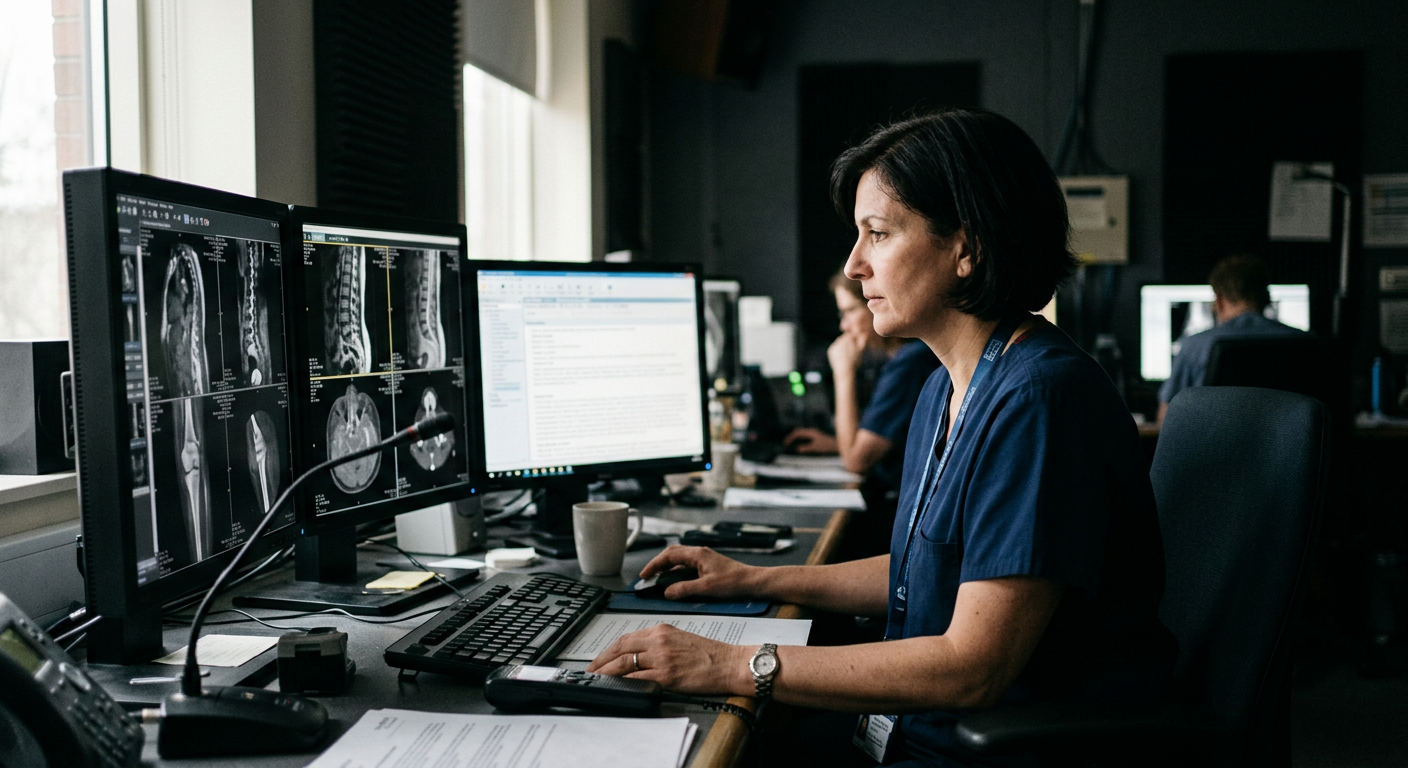
In clinical reading rooms, the traditional workflow of dictating a report from scratch is shifting to an editorial review process. When a computed tomography scan loads, a pre-written draft of the findings appears immediately, highlighting potential fractures and anomalies. This represents a fundamental change in how clinical documentation is produced.
On November 11, Mosaic Clinical Technologies, a subsidiary of the national practice Radiology Partners, reported results from an ongoing internal study of its investigational vision-language AI models. Developed by the organization's Cognita Imaging AI lab, these models draft complete radiology reports. The retrospective analysis of 237,905 imaging exams suggests that these AI-generated drafts improve clinical detection rates while increasing diagnostic speed.
A Shift from Detection to Drafting
Traditionally, artificial intelligence in radiology has functioned as a computer-aided detection tool, highlighting specific regions of interest for human evaluation. The system developed by Cognita Imaging AI lab, known as Mosaic, takes a different approach by generating complete, narrative draft reports. This transitions the radiologist from the role of primary author to that of an editor who reviews, refines, and signs off on the generated text.
The retrospective study evaluated these vision-language models across 50 clinical conditions on both computed tomography and X-ray examinations. The generated drafts surfaced additional clinically relevant findings subsequently confirmed by blinded expert radiologists. On average, including the AI draft report improved the detection of these findings by approximately 40 percent across the entire dataset of 237,905 exams.
This approach targets the primary administrative bottleneck in modern medical imaging. Radiologists spend a substantial portion of their working hours typing and dictating findings. By presenting a structured draft immediately upon opening an exam, the workflow changes from generation to verification, potentially reducing cognitive fatigue and standardizing reporting formats across a large clinical practice.
Examining the Clinical Data
The study broke down performance metrics across different imaging modalities. The enhanced detection rate achieved by using the draft reports averaged 40.9 percent for computed tomography scans and 43.4 percent for chest X-rays. For other types of X-rays, the average improvement in detection was 23.9 percent.
Certain specific clinical findings saw even larger increases. For example, the detection of fractures on head computed tomography scans rose by 73.4 percent when utilizing the AI-generated drafts. Similarly, the identification of lung nodules or masses on chest imaging increased by 57.5 percent. In some cases, the improvement in detection for specific clinical findings reached as high as 110 percent.
In addition to accuracy, the study measured the error rates of the generated drafts. On chest X-rays, 91.2 percent of the AI-generated draft reports were entirely free of errors, compared to 83.0 percent of the reports authored solely by radiologists. This was accompanied by measurable improvements in workflow efficiency. Experienced radiologists read computed tomography scans more than 27 percent faster when utilizing the drafts, while reading speed for X-rays improved by 31 percent.
Scaling to Eight Million Exams
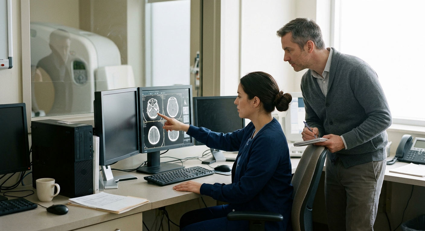
The clinical utility of these models is being tested on a large scale. By October 22, the Cognita Imaging AI lab models had drafted nearly 8 million exams in total, with the system currently processing more than 37,000 daily exams. This high volume provides a large dataset for ongoing evaluation under research protocols.
Despite the scale of its current deployment, the technology remains investigational. The models operate under a research protocol designed to gather the clinical evidence necessary to support official Food and Drug Administration authorization. The project has already received a Breakthrough Device designation from the Food and Drug Administration for the chest X-ray model in March.
The sheer volume of exams processed daily highlights the substantial infrastructure required to run large-scale clinical AI. Managing 37,000 daily drafts requires seamless integration with existing picture archiving and communication systems and radiology information systems across multiple facilities.
Workflow Integration and Market Read
The results represent a proprietary milestone for Radiology Partners. Because the company controls both the underlying technology through its subsidiary and the massive imaging volume of its national practice, it possesses the clinical workflow to test and deploy these models at a scale that few independent software developers can match.
However, the current findings are based on an internal, company-run study that has not yet undergone peer review. Independent validation will be an important step in establishing the broader clinical credibility of the system. For healthcare operators, the primary question is whether detecting a higher volume of findings translates directly to better clinical outcomes. There is also a practical concern regarding whether increased detection rates might lead to a rise in unnecessary follow-up imaging, which could add costs and anxiety for patients without offering a clear therapeutic benefit.
Source: The HealthTech Signal