
Listen instead
A physician at a workstation rotates a digital three-dimensional reconstruction of a patient's left anterior descending artery. With several clicks, the operator tests different stent lengths and diameters, measuring simulated blood flow improvements before the patient ever enters the catheterization suite. This shift from reactive, real-time decision-making inside the blood vessel to noninvasive, pre-procedural planning is moving from pilot programs into mainstream clinical practice. The transition is driven by a growing body of clinical evidence suggesting that preparing the procedural roadmap on a screen before making an incision can match the safety of invasive imaging while streamlining hospital operations.
Noninvasive Planning Proves Its Worth
At the Transcatheter Cardiovascular Therapeutics conference in October 2024, researchers presented data that challenges how clinicians approach percutaneous coronary intervention, or PCI. The investigator-initiated P4 trial, published in the New England Journal of Medicine, demonstrated that noninvasive CT-guided planning is clinically noninferior to intravascular ultrasound, known as IVUS, which has long served as the standard invasive imaging modality for complex cases.
The trial randomized 1,105 patients with stable coronary disease across 23 clinical centers in the United States, Europe, and Japan. Half of the cohort underwent PCI planned with coronary CT, while the other half received conventional guidance using IVUS during the catheterization. At 12 months, the rate of target-vessel failure was 4.1 percent in the CT-guided planning group compared to 4.6 percent in the IVUS-guided group. This met the statistical criteria for noninferiority, showing that pre-procedural planning with computational software yields clinical outcomes comparable to invasive, real-time vessel imaging. For clinical teams, this provides reassurance that omitting the invasive ultrasound catheter does not compromise patient safety.
Inside the Numbers

To plan the procedures, physicians in the CT arm used Heartflow's FFR-CT Analysis and the Heartflow Planner software. This technology integrates standard coronary CT imaging with anatomical and physiological analysis to assist in stent selection before catheterization begins.
The trial data showed clear operational differences between the two methods. The median procedure time for CT-planned cases was 46 minutes, compared to 63 minutes for the IVUS-guided cases, representing a 17-minute reduction in catheterization lab occupancy. Additionally, the median volume of contrast dye used was lower in the CT group at 150 milliliters versus 170 milliliters in the IVUS group. Calcium modification strategies were used in 9.2 percent of cases in the CT arm compared to 3.8 percent in the IVUS arm, indicating that the pre-procedural CT scans allowed operators to anticipate calcified lesions and prepare specialized tools in advance.
According to the study investigators, the median age of the trial participants was 65, and approximately 22 percent of the enrolled patients were women. Dr. Carlos Collet, the co-principal investigator from the Cardiovascular Research Foundation, stated that the P4 trial shows that CT-guided PCI planning achieves comparable clinical outcomes with shorter procedure times and less contrast. This approach allows interventionalists to anticipate anatomical challenges and tailor their strategy before the invasive procedure begins.
Shifting the Value Proposition
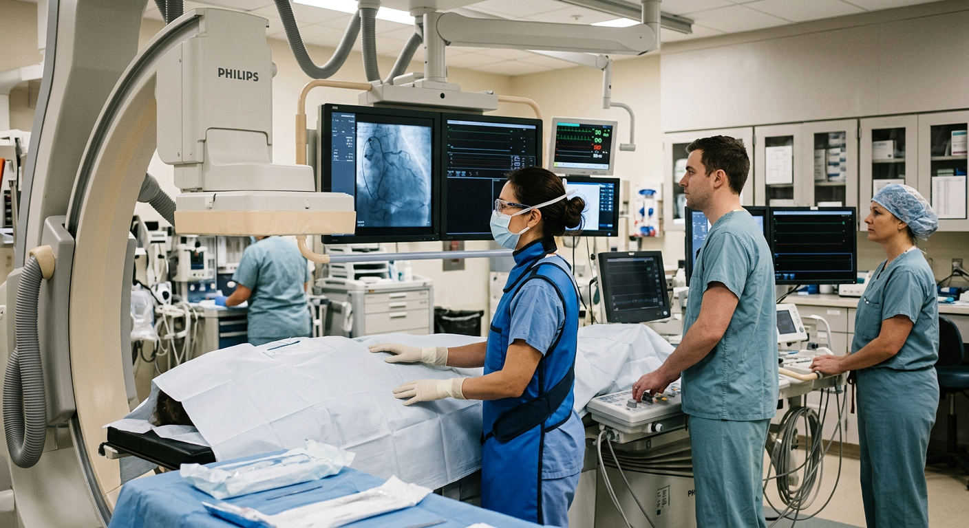
The clinical implications of the P4 trial are supported by separate findings from the NAVIGATE-PCI registry. In that 203-patient registry, physicians reviewed their treatment plans using the PCI Navigator software and compared them to angiography alone. After reviewing the software analysis, physicians altered their clinical plan in 70 percent of patients. The most frequent modifications involved adjusting the planned stent length in 41 percent of cases and altering the lesion preparation strategy in 12 percent of cases.
For decades, IVUS has been favored in interventional cardiology because visualizing the vessel wall from the inside improves patient outcomes by ensuring optimal stent expansion. However, performing IVUS adds time, material cost, and technical complexity to the procedure itself. The P4 trial suggests that much of the clinical planning can be completed before the patient is on the table. This transition shifts the economic and clinical value from invasive catheterization tools to diagnostic CT scanners and the software that interprets their images. Heartflow, which funded the P4 trial, reports that its technology is currently used at more than 1,800 institutions worldwide, suggesting that the infrastructure for this workflow shift is already partially established.
Operator and Economic Hurdles
While the trial results are promising, several operational questions remain for hospital administrators and clinical directors. The primary challenge is reimbursement. Healthcare systems must determine who pays for pre-procedural planning software and whether those costs can be offset by the savings from reduced catheterization lab time. A 17-minute reduction in lab time per procedure is operationally meaningful, as catheterization suites are among the most expensive assets in a hospital, often billed at high minute-based rates. Saving this time can allow high-volume centers to add additional procedures to their daily schedules.
Additionally, further research is required to determine if these results hold true for highly complex coronary lesions, such as chronic total occlusions, and if they can be replicated in community hospitals that lack specialized coronary CT programs or dedicated cardiac imagers. Hospitals will need to train technologists and cardiologists to interpret these pre-procedural maps effectively to realize the full efficiency gains demonstrated in the trial.
Source: The HealthTech Signal