Listen instead
Rhys Hibbert lay on an operating table at the National Hospital for Neurology and Neurosurgery in London while a surgical team prepared to navigate a path through his nasal cavity to reach an 11-millimeter pituitary tumor. The mass was pressing against his optic nerves, threatening his vision. To remove it safely, the surgeons had to guide their instruments within millimeters of major cerebral arteries and the optic chiasm. In this highly confined space, even a minor deviation can cause permanent blindness, stroke, or fatal hemorrhaging. Instead of relying solely on their own eyesight and static pre-operative scans, the surgeons used an active computer-vision system to analyze the live endoscopic feed during the procedure.
The operation represents a critical milestone in the clinical application of artificial intelligence. While machine learning has found a comfortable home in diagnostic imaging and administrative triage, its deployment as a real-time guide during active, invasive surgery has remained largely experimental. By deploying the system during a live pituitary resection, the clinical team demonstrated how computer vision can function as an active safety layer, identifying anatomical boundaries at the exact moment a surgeon is making critical cuts.
Computer vision in the operating room
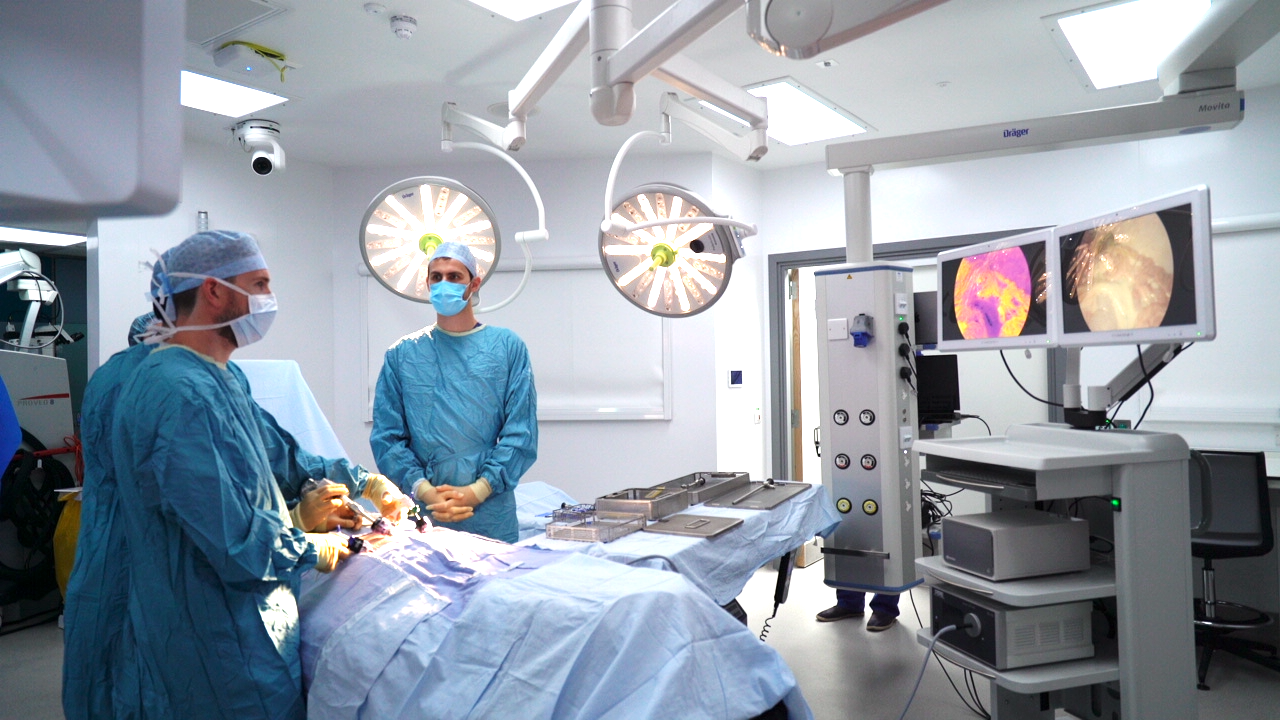
The procedure, performed by Professor Hani Marcus and neurosurgical registrar Danyal Khan, relied on a customized deep-learning model designed to identify anatomical landmarks on a live video feed. As the endoscope moved through the nasal cavity toward the skull base, the system processed the video stream in real time, projecting colored overlays onto the surgical monitor to highlight the internal carotid arteries, the optic nerve, and the boundaries of the pituitary gland itself.
This real-time visualization directly supported the surgical decision-making process. The tumor was successfully removed, and the surrounding critical structures remained entirely undamaged. Upon recovering from anesthesia, the patient reported an immediate improvement in his vision. Within a week of the procedure, he was discharged and walking independently.
For neurosurgery, where the margin for error is measured in fractions of a millimeter, the trial provides concrete evidence that real-time computer vision can be integrated into a standard surgical workflow without introducing distracting delays or technical friction.
The shift from static scans to live tracking

Historically, surgical navigation has relied on image-guided surgery systems that function much like GPS, mapping physical instruments to pre-operative magnetic resonance imaging or computed tomography scans. While useful, this approach has a fundamental vulnerability: it relies on static images. Once an operation begins, tissue shifts, fluids accumulate, and the physical reality inside the patient changes. A pre-operative scan cannot show the surgeon how the carotid artery has moved in response to the removal of surrounding bone or tumor tissue.
To bridge this gap, researchers at University College London trained their model on annotated endoscopic video data from hundreds of previous pituitary surgeries. By teaching the algorithm to recognize visual cues, such as the distinct texture and color of specific tissues under endoscopic light, they created a system capable of interpreting the dynamic operating environment.
This shift from spatial registration based on old scans to semantic understanding based on live video represents a major technical evolution. The system does not need to align the patient to a pre-operative coordinate space; instead, it looks at what the surgeon looks at and applies its training to label the structures in view.
Hardware, funding, and clinical integration
Processing high-definition surgical video and rendering analytical overlays with zero perceivable delay requires significant computational power. To achieve the low latency necessary for safe clinical use, the system was deployed on the NVIDIA Clara IGX platform. This specialized hardware architecture is designed specifically for medical edge computing, providing the high-throughput processing needed to ensure the AI's visual feedback matches the surgeon's physical movements in real time.
The development and clinical trial of the technology were supported by a coalition of public and private entities, including the National Institute for Health and Care Research, the Wellcome Trust, the Royal College of Surgeons of England, the Engineering and Physical Sciences Research Council, and Google.
This diverse funding coalition highlights the complex ecosystem required to transition healthcare AI from academic research to the operating room. The involvement of major academic centers, state health systems, and technology providers is becoming the standard template for validating high-stakes clinical software.
Clinical governance and the regulatory pathway
From an industry perspective, the design of this trial offers a masterclass in navigating clinical risk and regulatory pathways. The AI system did not control any surgical instruments, nor did it automate any physical movements. It functioned entirely as an advisory tool, offering a digital safety net by highlighting structures that might otherwise be obscured by blood, bone dust, or tumor debris.
By keeping the human surgeon in absolute control of every physical action, the developers bypassed the most daunting regulatory and liability hurdles associated with autonomous medical systems. The clear division of labor keeps clinical responsibility squarely with the credentialed surgeons.
This pragmatic approach is likely to accelerate the commercial adoption of computer vision in operating rooms. Rather than waiting for the resolution of complex legal and ethical debates regarding autonomous robotic surgery, medical device companies and health systems can deploy assistive, vision-based tools today. These systems improve safety, reduce complications, and collect the massive datasets required to train the autonomous systems of the future, all while operating safely within current regulatory and liability frameworks.
Source: University College London Hospitals NHS Foundation Trust